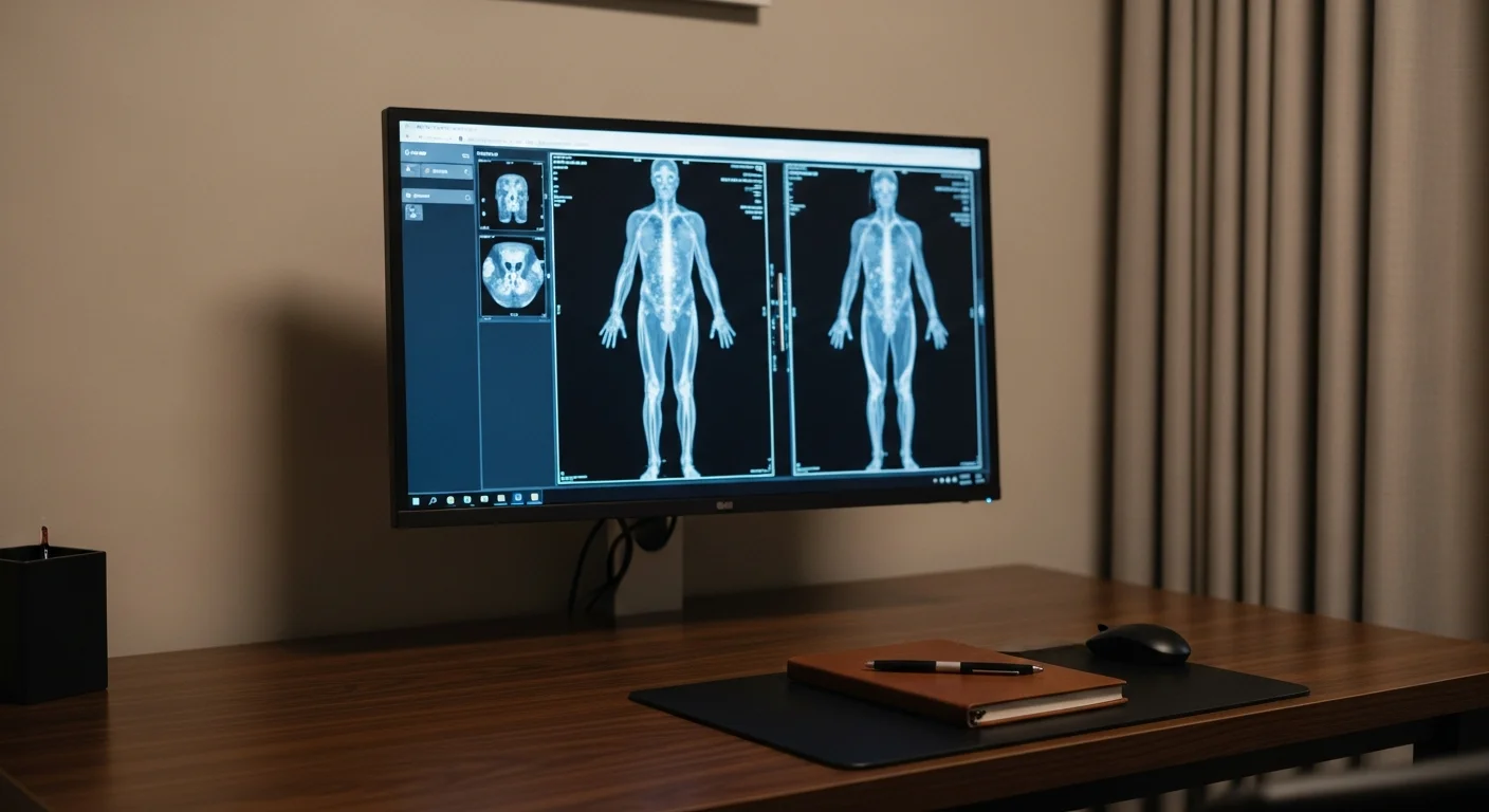
MRI uses a magnetic field rather than radiation to produce the most detailed view available of soft tissue, which makes it the study for the brain and spinal cord, cartilage and ligaments, bone marrow, and organs where the question is tissue character rather than shape. Fishtown Medicine orders MRI when a specific question needs that detail, not as a broad look, because degenerative findings appear on the scans of most pain-free adults and an early MRI often adds findings without adding answers.
TL;DR: MRI gives the most detailed picture of soft tissue that medicine has, with no radiation at all, and that combination makes it feel like the study everyone should get. The catch is that detail is not the same as an answer. Degenerative findings turn up on the spine MRIs of most adults who have no pain whatsoever, so a scan ordered before the clinical picture is clear tends to produce a list of abnormalities that may have nothing to do with why you hurt. The right time for an MRI is when there is a specific question it can settle: a nerve problem with weakness, a knee that gives way after failed therapy, a mass that needs its tissue characterized, or anything involving the brain or spinal cord. It takes 20 to 60 minutes, it is loud, and metal in your body has to be sorted out beforehand rather than at the scanner door.
What does an MRI show that a CT or X-ray cannot?
MRI works on a different principle from the other studies. Rather than passing radiation through you and measuring what gets absorbed, it places you in a strong magnetic field, nudges the hydrogen atoms in your tissues with radio waves, and listens to the signal they give off as they settle back. Because hydrogen sits mostly in water and fat, and because different tissues hold water differently, the machine can distinguish tissues that an X-ray beam treats as nearly identical.
That is the whole advantage. An X-ray separates bone from everything else. A CT separates bone, fat, fluid, and tissue reasonably well and does it fast. An MRI separates grey matter from white matter, cartilage from the bone beneath it, a ligament from the fluid around it, a tumor from the swelling surrounding it, and healthy marrow from marrow that is inflamed or infiltrated. It also does this in any plane without moving you, and it can be tuned with different sequences to emphasize whatever the question requires.
The tissue where this matters most is the nervous system. The brain and spinal cord are nearly invisible on plain film and only roughly rendered on CT, so any question about multiple sclerosis, a spinal cord problem, a pituitary abnormality, a seizure focus, or the cause of new neurologic symptoms is an MRI question. Bone marrow is the second place MRI stands alone, which is why it finds a stress fracture in the first week, when the X-ray still looks normal, and why it is the study for a suspected bone infection.
When is an MRI the wrong first study?
The strongest argument against an early MRI is not cost or access. It is that scans of healthy people are full of abnormal-looking findings.
A systematic review pooling 33 studies of 3,110 people with no back pain at all found disc degeneration in 37% of 20-year-olds rising to 96% of 80-year-olds, disc bulges in 30% at age 20 rising to 84% at age 80, and disc protrusions in 29% at age 20 rising to 43% at age 80.1 These were people who felt fine. The authors concluded that many of these features are part of ordinary aging rather than a source of pain.
Sit with what that means for a person with back pain who gets an early MRI. The scan will very likely show something. That something has perhaps a 1 in 3 chance of being present in a pain-free person the same age. Now you and I have a finding on a report, a word like "protrusion" attached to your spine, and no reliable way to know whether it explains your symptom. That uncertainty tends to lead toward injections and surgical consultations rather than toward relief, and it is why the imaging strategy trials found that early imaging did not improve pain or function.
So MRI belongs after the clinical picture narrows the question, not before. For back pain that means surgical planning, a progressive neurologic deficit, or pain past 6 weeks that has fully failed physical therapy. For a knee it means instability that persisted through rehabilitation, or a clear traumatic event. For a shoulder it means a suspected full-thickness tear after therapy has been tried. The exception in every region is a red flag, since suspected infection, cancer, cord compression, or cauda equina goes to MRI immediately.
When do I need contrast, and is gadolinium safe?
Many MRIs are done without any contrast at all, and for musculoskeletal questions such as a meniscus, a rotator cuff, or a disc, the plain study is usually all that is needed.
Contrast is added when the question is about blood supply or an active process. Gadolinium is a metal that alters the magnetic behavior of nearby tissue, and it leaks out of blood vessels wherever they are abnormally permeable, which is what happens around tumors, infections, active inflammation, and areas where the blood-brain barrier has broken down. So a search for a brain tumor, a suspected abscess, an assessment of multiple sclerosis activity, or a characterization of a liver or kidney mass usually means contrast. Cancer follow-up almost always does.
On safety, the main historical concern was nephrogenic systemic fibrosis, a serious condition seen in people with severe kidney impairment exposed to older gadolinium agents. Practice changed in response: kidney function is checked before contrast in anyone at risk, and the newer, more stable agents now in routine use carry a very low risk even in advanced kidney disease. The other topic you may have read about is gadolinium retention, meaning trace amounts detectable in tissue years later. Retention is measurable; a resulting illness in people with normal kidneys has not been established. What I want you to know is that this is a genuine trade rather than a settled non-issue, which is why contrast gets added when it answers something and left off when it does not.
Tell the technologist if you are pregnant, breastfeeding, have kidney disease, or have had a reaction to contrast before.
What is the scan like?
An MRI takes 20 to 60 minutes depending on the region and how many sequences are needed, and it requires you to hold still for the whole thing, since motion blurs the images in a way that can force a repeat.
It is loud, because the gradient coils that build the image knock and buzz at volumes that need ear protection, which the facility provides. You lie on a table that slides into a tube, and how enclosed you feel depends on what is being scanned, since a knee study may leave your head outside the bore while a brain study puts a coil close around your face.
Claustrophobia is common and worth raising in advance rather than discovering at the scanner. There are several workable answers: a short course of an oral anxiolytic taken before the appointment with a driver arranged, a wide-bore scanner that is noticeably roomier, an open MRI when the question tolerates lower image quality, and simple measures such as an eye mask, a mirror that lets you see out of the bore, or having someone stay in the room with you. Nobody should white-knuckle it, and nobody should skip a study they need because the first appointment was frightening.
Fishtown Medicine
A 90-minute conversation with Dr. Ash. A written plan you can actually follow.
Metal is the part to sort out early. Pacemakers and implanted defibrillators, cochlear implants, certain aneurysm clips, some neurostimulators, and any history of metal fragments in the eye all require review before scheduling, and some are absolute contraindications. Most modern orthopedic hardware, joint replacements, and dental work are safe, though hardware near the area of interest can distort the picture. Bring the card or the model number if you have one, since MRI safety is determined by the specific device rather than the category.
How does MRI compare with CT for the same body part?
For the brain, CT is the emergency study and MRI is the detailed one. CT takes seconds and is excellent at fresh bleeding and skull fracture, which is why it is what happens after head trauma or a suspected acute stroke. MRI shows the brain tissue itself far better and is the study for anything subacute or chronic. The brain imaging guide works through that split in more depth.
For the spine, X-ray shows alignment and bone, and MRI shows discs, nerve roots, and the cord. There is no overlap worth arguing about; they answer different questions.
For joints, X-ray answers fracture and arthritis, and MRI answers cartilage, ligament, meniscus, and labrum. Most joint pain starts with the plain film and stops there, which the X-ray guide covers body part by body part.
For the abdomen, CT is faster, cheaper, and usually first, while MRI is better at characterizing a liver lesion, assessing the bile ducts, or working up a pancreatic question when CT has left ambiguity.
For screening a person with no symptoms, whole-body MRI is a category of its own with a specific problem attached, which the boutique scan review addresses directly.
Guidance from the Clinic

How Fishtown Medicine decides on an MRI in Philadelphia
The question I work through before ordering one is what I would do differently depending on the result. If the honest answer is that physical therapy comes next either way, the scan waits. If a specific finding would change the plan, whether that means a surgical referral, a different diagnosis, or stopping a treatment that is not addressing the problem, it is worth doing.
When an MRI is the right study, Fishtown Medicine arranges it rather than handing over an order. That includes checking that implants are cleared beforehand, sorting out claustrophobia in advance, and pointing self-pay patients toward independent imaging centers in the Philadelphia area, where the same study on comparable equipment often costs a fraction of the hospital-affiliated price.
Results come with a conversation. An MRI report is dense, it lists everything the radiologist sees whether or not it matters, and read alone it is frightening. What you need is which finding explains your symptom, which findings are ordinary for your age, and what happens next.
Key Takeaways
- MRI uses magnetism rather than radiation and gives the most detailed soft-tissue picture available, which makes it the study for the brain, spinal cord, cartilage, ligaments, and bone marrow.
- Detail is not the same as an answer. Disc degeneration appears in 37% of pain-free 20-year-olds and 96% of pain-free 80-year-olds, so an early scan often adds findings rather than clarity.
- The right moment is when a specific finding would change the plan: surgical planning, a progressive neurologic deficit, failed conservative care, or any red flag.
- Contrast is added when the question involves blood supply or an active process such as tumor, infection, or inflammation, and it is left off when it would not change the answer.
- Sort out implants and claustrophobia when scheduling, not at the scanner. Device safety depends on the specific model.
- MRI finds a stress fracture in week 1 because it sees marrow swelling directly, while an X-ray stays normal until healing bone appears.
Related at Fishtown Medicine
- When You Need an X-Ray - the first study for most bone and joint questions
- When You Need an Ultrasound - the radiation-free option for superficial structures and flow
- Understanding Brain and Head Imaging - how CT and MRI divide neurologic questions
- Bone and Joint Imaging 101 - the musculoskeletal sequence in detail
- Boutique Scans and Whole-Body MRI - why screening a person without symptoms is a different problem
- When to Order Imaging - the decision framework behind every scan
Scientific References
- Brinjikji W, Luetmer PH, Comstock B, et al. "Systematic literature review of imaging features of spinal degeneration in asymptomatic populations." AJNR American Journal of Neuroradiology. 2015;36(4):811-816. PubMed
- Chou R, Fu R, Carrino JA, Deyo RA. "Imaging strategies for low-back pain: systematic review and meta-analysis." Lancet. 2009;373(9662):463-472. PubMed
- Mettler FA Jr, Huda W, Yoshizumi TT, Mahesh M. "Effective doses in radiology and diagnostic nuclear medicine: a catalog." Radiology. 2008;248(1):254-263. PubMed

Frequently Asked Questions
Common Questions
Deep-Dive Questions
Ready when you are
Dr. Ash reads every intake himself, and answers questions personally - usually within a few hours.